CHAPTER3
Movement
Have you ever marveled at the athleticism of a tennis player as she lands a perfect serve, or the virtuosity of a pianist whose fingers dance through a piece by Rachmaninoff? These are special and dramatic movements. Yet in our daily lives, each of us performs a suite of complex, skilled movements that are equally remarkable — from walking and talking, to signing our names, or sending a text. We even use our muscles to reveal our current mood: A smile and a wave are universally understood.
Movement is such an integral part of our day-to-day experience that we take for granted the sophisticated systems that make these actions possible. The central nervous system — brain and spinal cord — directs the coordinated actions of the hundreds of muscles that enable us to move. These actions are refined and strengthened as we make our way through the world, adapting to changing circumstances and practicing, sometimes even improving, our motor skills.
VOLUNTARY MOVEMENTS
 To understand how the nervous system governs motion, we begin with the muscles, the structures of the body that produce movement. Most muscles attach to the skeleton and span joints, the sites where two or more bones come together. The close relationship of these muscles to the skeleton gives them their name — skeletal muscles. Activating muscles can either flex or extend the joint that they span. Muscles that bend a joint, bringing the bones closer together, are called flexors; muscles that straighten the joint, increasing the angle between the bones, are called extensors. Flexors and extensors work in opposition, so when one set of muscles contracts, the other relaxes. For example, bending the elbow requires contraction of the biceps (a flexor) and relaxation of the triceps (an extensor). For such motions, the muscles that promote the movement are called agonists, and those that oppose or inhibit the movement are antagonists. Skilled, rapid movements — like throwing a dart — are started by agonists and stopped by antagonists, allowing the limb to accelerate and halt with great speed and precision. For some movements, agonists and their opposing antagonists contract at the same time, which is called co-contraction. These simultaneous actions can stabilize or control a movement, such as holding an object at arm’s length or stabilizing an immobile joint during isometric exercises.
Whether flexion or extension, the movement of all skeletal muscles is controlled by the central nervous system. A skeletal muscle is made up of thousands of individual muscle cells, called muscle fibers. Each muscle fiber is controlled by a single alpha motor neuron that originates in the spinal cord or the brain. However, each of these alpha motor neurons can control multiple muscle fibers (from a few to 100 or more). An alpha motor neuron plus all the muscle fibers it controls form a functional unit known as a motor unit, the critical link between the central nervous system and skeletal muscles. When motor neurons die — as happens in diseases like amyotrophic lateral sclerosis (ALS) — people can lose their ability to move.
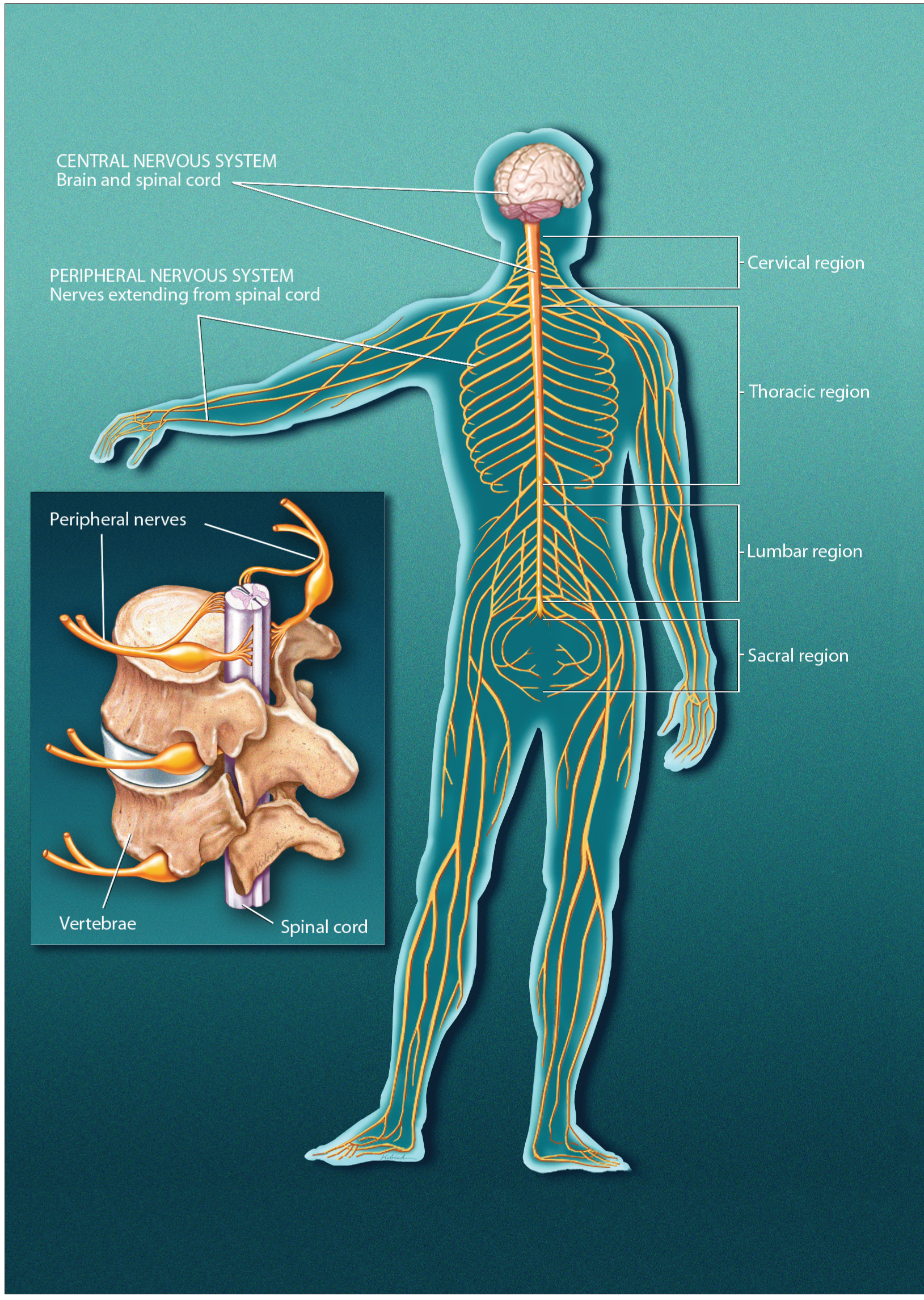
The nervous system is divided in two. The central nervous system consists of the brain and spinal cord. The peripheral nervous system consists of nerves and small concentrations of gray matter called ganglia. The brain sends messages to the peripheral nerves, which control the muscles and internal organs.
Some muscles act not on joints but on soft tissue. For example, muscles in the head and neck enable us to move our eyes, chew and swallow food, have conversations, and control our facial expressions. These muscles are also controlled by the central nervous system, and they operate in much the same way as those that attach to bones.
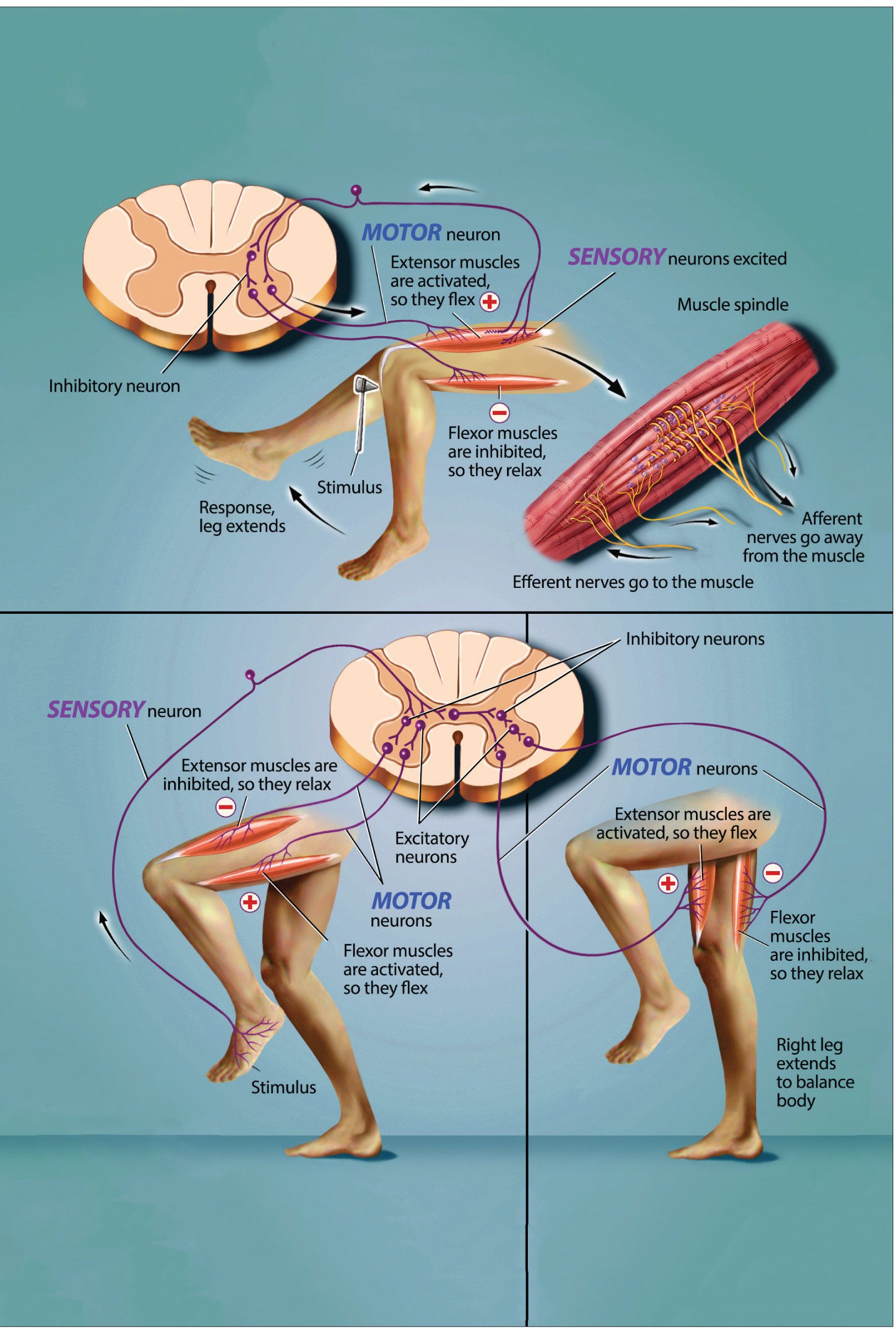
The stretch reflex, as seen at top of the image, occurs when a doctor taps a muscle tendon to test your reflexes. This activates muscle spindle sensory fibers, which send a barrage of impulses to the spinal cord, activating motor neurons and triggering muscle contraction. Flexion withdrawal, shown on the bottom of this image, occurs when you step on a sharp object, and your leg is immediately lifted (flexion) from the source of potential injury. The opposite leg responds with increased extension so that you can maintain your balance, called the crossed extension reflex.
INVOLUNTARY MOVEMENTS
Many types of movement take place without our conscious control. Among the simplest and most fundamental types of involuntary movements are the reflexes. Reflexes are relatively stereotyped, automatic muscle responses to particular stimuli — think of the rapid withdrawal of your hand after touching something hot. These reflexes involve the activation of sensory receptors in the skin, the joints, or even in the muscles themselves. The responses are rapid and occur without involvement of the brain or conscious attention. Instead, they depend on circuits of neurons located in or near the spinal cord itself.
One of the best-known reflexes is the “knee jerk” response, a stretch (myotatic) reflex that occurs when a physician strikes the tendon just below the knee with a small rubber hammer. This tap produces a slight stretch of the knee extensor muscle, which is “sensed” by receptors within the muscle called muscle spindles. The spindles sense the extent and speed of the stretch, and stimulate sensory neurons, which send a barrage of impulses into the spinal cord. There, the signals activate the alpha motor neurons that cause the stretched extensor muscle to contract, triggering the reflex. Of course, for the leg to kick forward, the antagonist flexor muscle has to relax at the same time. In fact, the same sensory stimulus that directly activates the motor neurons controlling the extensor also indirectly inhibits the motor neurons controlling the antagonist flexor. This reciprocal inhibition is accomplished by connecting neurons that lie completely within the spinal cord. When these so-called inhibitory interneurons are activated by the original sensory stimulus, they send impulses that inhibit the motor neurons supplying the flexor.
Thus, even the simplest of reflexes involves the synchronous activation (and inactivation) of multiple sets of motor neurons controlling both agonist and antagonist muscles.
Many reflexes protect you from injury. When you’re seated in a doctor’s office, the “knee jerk” reflex simply makes your lower leg swing briefly forward. However, if you were to jump off a chair (or perform an even more dramatic gymnastic dismount) this same reflex would promote the contraction of the strong muscles that straighten your knees, helping you to “stick your landing” and remain upright. Another protective reflex is the flexion withdrawal reflex that occurs when your bare foot encounters a sharp object. In this case, pain receptors in the skin send a message to the spinal cord, alpha motor neurons are activated, and the leg is immediately lifted (flexion). At the same time, because your body weight is supported on both legs, the extensors of the opposite leg must be activated. Without this additional reaction, called the flexion crossed extension reflex, you would lose your balance and fall over after stepping on a tack.
Fallini, et al. The Journal of Neuroscience, 2016.
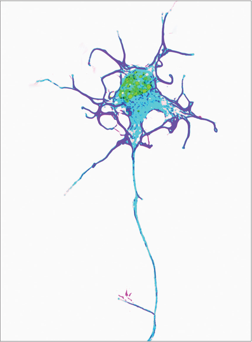
Specialized cells called motor neurons carry instructions from the brain along long axons that stretch from your spinal cord to the muscles in your hands and feet. These cells can be the longest in your body, stretching the length of your leg to control the muscles in your feet.
Wright, et al. The Journal of Neuroscience, 2009.
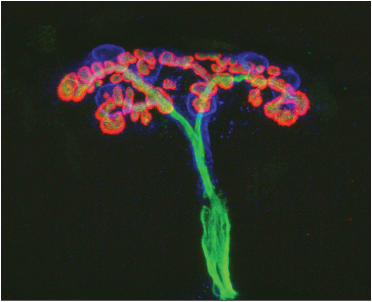
Neurons communicate with muscles at sites called neuromuscular junctions. This image shows a neuromuscular junction in a mouse, with a motor neuron labeled in green and neurotransmitter receptors on muscle cells labeled in red.
As all these movements occur, the muscles involved provide feedback to the brain with information about where the various body parts are in space and how fast they are moving. The muscle spindles mentioned earlier supply information about changes in muscle length or stretch. The brain, in turn, adjusts the sensitivity of the system via a separate set of motor neurons, gamma motor neurons, which keep the muscle spindles taut. Other specialized receptors called Golgi tendon organs — located where the muscle fibers connect to the tendon — detect how much force or tension is applied to a muscle during ongoing movement, increasing the movement’s precision. These feedback systems are not unique to reflexes, but allow the brain to fine-tune how working muscles behave during a variety of movement tasks — from those that require a mastery of delicate positioning and coordination, such as sipping from a dangerously full teacup, to those that involve a targeted application of strength and speed, such as throwing a runner out at first base.
The most complex movements that you perform, including those requiring conscious planning, involve input from the brain.
VOLUNTARY AND COMPLEX MOVEMENTS
Spinal circuits also play a critical role in controlling more sophisticated, voluntary behaviors, such as the alternating action of the legs during walking. In fact, the rhythmic patterns of muscle activation that produce locomotion — not only in four-footed animals, but in humans — are generated by neurons within spinal cord and brainstem circuits. When these neuronal circuits (central pattern generators) are activated, they produce the rhythmic patterns that occur in walking, flying, swimming, or breathing. Central pattern generators which evolved in primitive vertebrates, are being studied to determine the degree to which spinal circuitry can be co-opted to recover basic postural and locomotor function after severe paralysis.
The most complex movements that you perform, including those requiring conscious planning, involve input from the brain. These higher brain regions initiate voluntary motion, coordinate complex sequences of movement, and tailor behavioral output to suit a given situation. Successful execution of these programs requires your brain to relay commands to the appropriate spinal circuits.
Through careful animal experiments, scientists are just beginning to understand the coordinated series of interactions that take place among different brain regions during voluntary movement. One brain area essential for voluntary movement is the motor cortex. Neurons in the motor cortex send signals that directly control the activation of alpha motor neurons in the spine. Some of these cortical neurons control the movement of functionally related muscles in an individual body part, such as your hand or arm; such neurons are important for finely tuned motor skills. Other neurons in the motor cortex can direct the coordinated movement of a limb to a particular point in space — raising your arm in a defensive position or bringing a hand to your mouth to deliver a tasty morsel of food.
Regions that Modulate Voluntary Movement
 The motor cortex does not act alone in controlling complex or skilled voluntary movements. Several other brain regions participate in parallel circuits or “loops” to modulate motor control. These regions — including the basal ganglia, thalamus, cerebellum, and a large number of neuron groups located within the midbrain and brainstem — also influence the activity of motor neurons in the spinal cord. The basal ganglia themselves encompass two separate pathways. One appears to facilitate the desired motor program while the other suppresses unwanted, competing actions. Along with the thalamus, the basal ganglia share widespread connections with motor and sensory areas of the cerebral cortex, allowing these structures to monitor and adjust motor performance.
Dysfunction of the basal ganglia can lead to serious movement disorders. People with Parkinson’s disease experience degeneration of neurons in a brain region called the substantia nigra; these neurons relay signals to the basal ganglia using the neurotransmitter dopamine, a key chemical involved in motor control. Depletion of dopamine gives rise to the hallmark symptoms of Parkinson’s: tremor, rigidity, and in some cases, akinesia, an inability to move. In contrast, individuals with Huntington’s disease often display uncontrolled jerking or twitching movements, particularly in the face and extremities. These symptoms stem from a selective loss of inhibitory neurons in the basal ganglia, which eliminates the suppression of random involuntary movements.
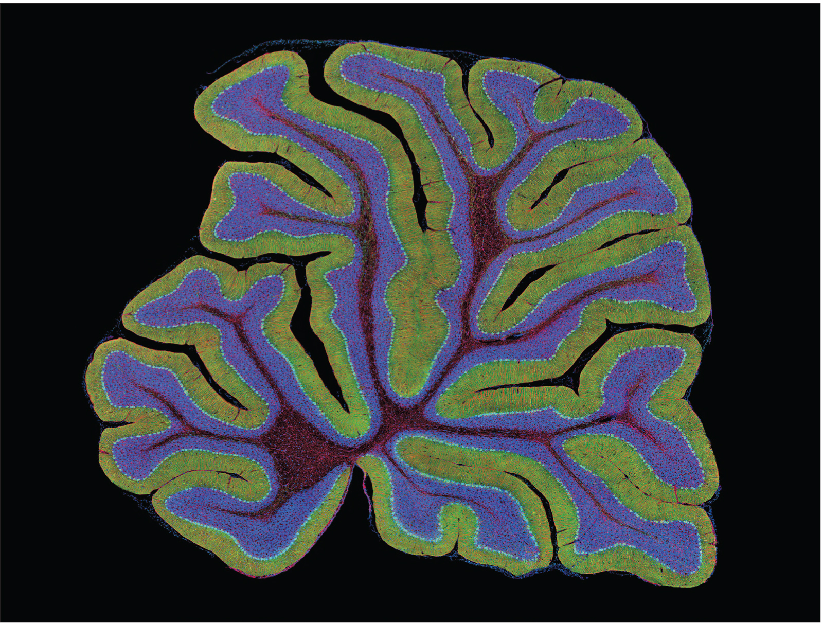
Thomas Deerinck, National Center for Microscopy and Imaging Research, University of California, San Diego.
The cerebellum, shown in this image of a mouse brain, is a region at the back of the brain associated with movement.
Another brain region crucial for coordinating and fine-tuning skilled movement is the cerebellum. The cerebellum receives direct input from sensory receptors in the limbs and head, as well as most areas of the cerebral cortex. Neurons in the cerebellum apparently integrate this sensory information ensuring the proper timing and integration of muscle action. This enables us to produce fluid movements more or less automatically. The cerebellum is essential to a wide range of motor learning and coordination, from controlling limb movements to eye movement to grip force.
Disturbance of cerebellar function leads to poor coordination, disorders of balance, and even difficulties in speech, one of the most intricate forms of movement control. Long-term alcohol abuse is a common cause of acquired cerebellar degeneration. Typical symptoms are poor coordination, an unsteady walk or stumbling gait, changes in speech, and difficulty with fine motor skills including eating, writing, and dressing.
The cerebellum also allows you to adapt to the unexpected, adjusting your movements so that you can smoothly lift a box that you expected to be much heavier, for example. And, it plays a major role in motor learning. As you learned to walk or speak or practiced a musical instrument or a new dance routine, the cerebellum refined and sharpened the motor programs that allow you to perform these tasks with increasing accuracy and skill.
Considerable evidence also indicates that the cerebellum helps us recalibrate our movements as our own bodies change, as we grow taller, gain or lose weight or muscle mass, or cope with disease or disability. In that way, the cerebellum facilitates skillful movement through an ever-changing world as we grow up and we grow old. 