CHAPTER1
Brain Basics
The brain is literally the “nerve center” of your body — it contains billions of neurons that transmit information from the body and the outside world, and then programs our responses — conscious and unconscious movements, thoughts, emotions, and memories. What’s more, your brain can do all these things simultaneously: You can throw a ball while talking to a friend, plan dinner while you’re shopping, or daydream about a balloon ride as you drive to work. Your brain can pull off these feats of multitasking because it is split into many distinct regions specialized for specific tasks and abilities.
Major Brain Landmarks
 The largest part of the human brain is the cerebrum. It is divided into two large, separate hemispheres, one on the left side, the other on the right. The hemispheres are connected by bundles of nerve fibers that carry information from one side of your brain to the other. The largest of these bundles forms a bridge between the cerebral hemispheres and is called the corpus callosum.
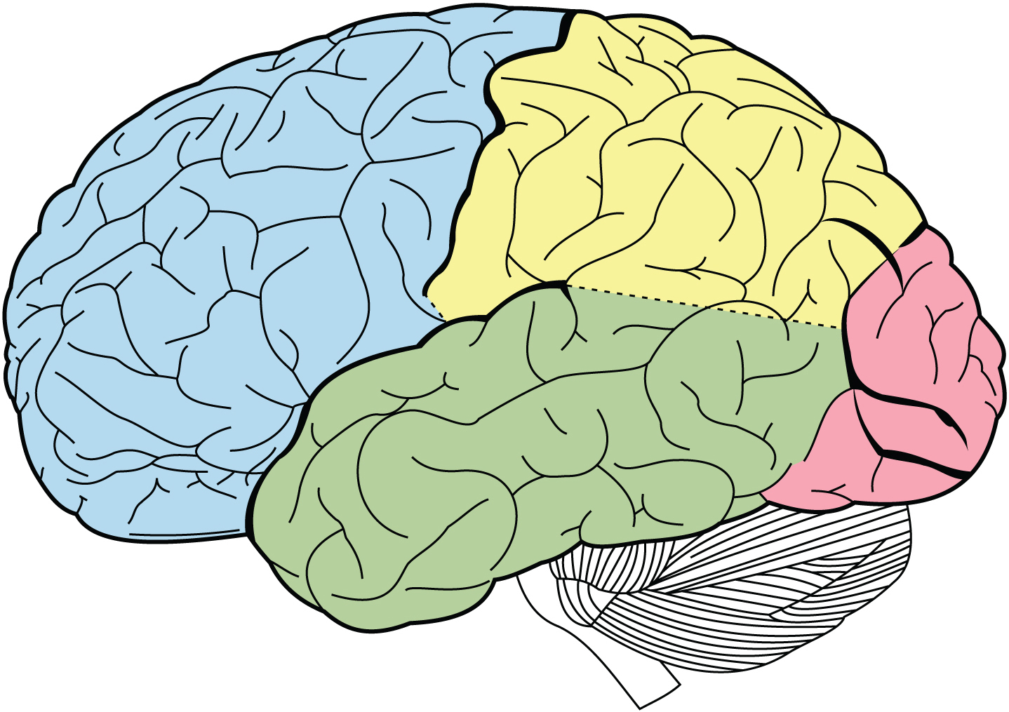
The surface of the cerebrum is a deeply folded layer of nerve tissue called the cerebral cortex. Its deep folds increase the area of the cerebral cortex, creating space in this surface layer for more neurons, which increase the brain’s processing power. Just as explorers use landmarks like rivers and mountain ranges to describe and map continents, neuroscientists use the deepest divisions of the cerebrum to identify regions of each hemisphere as separate lobes — distinct regions that have characteristic functions. This “brain map” will serve as a useful trail guide as you explore the brain in the chapters ahead.
The frontal lobes are at the front of the brain, immediately above the eyes. Parts of these lobes coordinate voluntary movements and speech, memory and emotion, higher cognitive skills like planning and problem-solving, and many aspects of personality.
The parietal lobes are located at the top of the brain, immediately behind the frontal lobes. They integrate sensory signals from the skin, process taste, and process some types of visual information.
The back of the brain houses the occipital lobes. They process visual information and are responsible for recognizing colors and shapes and integrating them into complex visual understanding.
The temporal lobes lie on the sides of the brain, at and below the level of the eyes. They carry out some visual processing and interpret auditory information. The hippocampus consists of curved structures lying beneath the cerebral cortex; it is a region of the temporal lobes that encodes new memories. Another deep structure within each temporal lobe, the amygdala, integrates memory and emotion.
The hippocampus and amygdala are part of the limbic system, a group of structures deep within the brain that help regulate our emotion and motivation. Other parts of the limbic system include the thalamus, which integrates sensory information and relays it to other parts of the brain, and the hypothalamus, which sends hormonal signals to the rest of the body through the pituitary gland. These structures, together with the cerebral cortex, make up the forebrain.
The midbrain sits beneath the thalamus. It includes distinct groups of neurons that coordinate eye movements like blinking and focusing, and trigger reflexes to sounds. An example is the startled jump when you are surprised by a loud noise. Other regions of the midbrain inhibit unwanted body movements and help coordinate sensory input and motor output to manage the fine motor control that enables you to write with a pen or play a musical instrument.
Henry Vandyke Carter
Pictured are the brain’s four principal lobes. The frontal lobe, responsible for attention, planning, and decision-making, is labeled blue. The temporal lobe, associated with language, memory, and emotion, is labeled in green. The parietal lobe, which integrates information from the senses, is labeled in yellow. And the occipital lobe, responsible for vision, is labeled in pink.
Some of these regions — along with parts of the forebrain — form a collection of structures called the basal ganglia, which helps regulate complex body movements.
The hindbrain plays roles in glucose regulation and sleep and includes several regions that help control movement. The cerebellum, tucked underneath the occipital lobe at the very back of the brain, is the second-largest part of the brain in volume, containing over half the brain’s neurons. Like the cerebrum, the cerebellum is deeply folded, divided into two hemispheres, and carries out a variety of functions. For example, it coordinates voluntary movements and helps the brain learn new motor skills. It also has roles in spatial and temporal perception. A patient with cerebellar damage might have a jerky, arrhythmic gait or might be unable to accurately touch his finger to his nose.
Below the cerebellum is the pons, which influences breathing and posture. Another part of the hindbrain, the medulla, carries nerve pathways connecting the brain to the spinal cord and contains neural networks that help control basic functions like swallowing, heart rate, and breathing. Together, the midbrain, pons, and medulla make up the brainstem.
Brain Evolution
It’s hard to believe that our complex human brain evolved from a simple tube. The earliest vertebrates probably had brains much like the one in the modern lancelet Amphioxus — little more than a wide spot in the hollow nerve cord running down its back. But while the lancelet’s brain looks simple, it still contains specialized regions where neurons process specific kinds of information, like the presence of light or the chemicals drifting through the water. In its early development, the human brain began as a simple tube, and even today it is divided into the same kinds of regions as the brains of our ancestors.
In early vertebrates, the “brain” end of the nerve cord developed three distinct bulges as neurons were added, improving processing in sensory and motor reflex regions. These bulges became the forebrain, the midbrain, and the hindbrain. In the forebrain, the region able to detect chemicals expanded to form the olfactory bulbs, and with the evolution of image-producing eyes, light-sensing regions expanded and began processing more complex visual signals. The cerebellum appeared as the hindbrain and expanded the regions that control escape movements and orient the body in space. Both these functions are far more important to an actively swimming fish than to a sedentary lancelet buried in the sand.
Regions that could rapidly process visual and auditory information and trigger appropriate escape, feeding, or mating behaviors also expanded in vertebrates. Over time, those new types of neurons made the forebrain balloon out, forming the cerebral hemispheres. In early mammals, cortical tissues in the cerebrum and the cerebellum expanded even further, packing new neurons into layers and folds generating more complex tissues with increased processing power.
NEURAL NETWORKS
 Information moves from one region of your brain to another via chains of neurons that can transmit signals over long distances. When the nerve fibers of region-spanning neurons form distinct bundles, these are called nerve tracts. Examples of major nerve tracts include the corpus callosum (the thick bundle of neurons connecting your left and right cerebral hemispheres) and the smaller anterior commissure that transmits signals between the left and right temporal lobes.
A group of nerve tracts connecting a series of regions in the brain is called a neural network. Neural networks route signals through the brain along a linear pathway, analyzing and organizing different types of information within fractions of a second.
Have you ever wondered what happens in your brain when you watch a movie? Your brain turns a panoply of moving shapes into recognizable characters and scenery. The process begins with photoreceptors, cells in the retina that trigger electrical signals in response to specific wavelengths of light. Once those signals reach the optic nerve, they travel through the optic tract to the thalamus, where neurons respond to the shape, color, or movement of objects on the screen and pass their signals to the primary visual cortex in the occipital lobe, at the back of the brain. Neurons in the primary visual cortex, in turn, detect the edges of objects within the field of vision and integrate the signals from each eye, creating a three-dimensional representation of the outside world. The image is even further refined as signals are sent down two parallel processing streams. In one stream, neurons in the temporal lobe recognize and identify objects; in the other, neurons in the parietal lobe detect the spatial location of those objects. And that’s only the visual input from the film! New technologies that allow us to look with increasing detail at the brain regions being activated as we perform different functions are giving us increasing insight into the fine regions of the brain used for specific tasks.
Network Activity Creates Brain Waves
The visual cortex also sends signals back to the thalamus to become integrated with other sensory information; this is an example of a “thalamocortical loop,” a two-way circuit that connects the thalamus with parts of the cortex and back. As neuronal signals loop through the thalamus and cortex, they produce rhythmic, oscillating, electrical patterns that can be detected with an electroencephalograph (EEG). These signals are commonly called brain waves. There are four distinct types, each recognized by its characteristic shape on an EEG display or printout.
Your awake brain typically produces alpha waves and beta waves. Alpha waves originate mainly in the parietal and occipital lobes when your brain is relaxed and eyes are closed, and are characterized by frequencies between 8 and 13 Hz. (The Hertz is a measure of frequency; 1 Hz = 1 cycle per second.) Beta waves are somewhat faster, with frequencies ranging from 14 to 30 Hz. Beta waves are typically produced by the frontal and parietal regions of your brain when it processes sensory input or concentrates on a task. Theta waves and delta waves are typical of sleep. Theta waves are slower than alpha waves, ranging from 4 to 7 Hz, while delta waves, which occur during deep sleep, are very slow, with frequencies less than 3.5 Hz. Alpha and delta waves are typically of higher amplitude (stronger) than beta or theta waves but, when measured with electrodes on your scalp, all these signals are in the microvolt range: 20–200 μV for alpha and delta waves, and 5-10 μV for beta and theta waves.
Neurons are organized into a stack of distinct layers that span the thickness of the cortex like shelves in a bookcase.
Neural Networks Organize and Integrate Information
Your brain and spinal cord contain many distinct neural networks. These include spinal tracts — chains of neurons that pass signals through the brainstem and the spinal cord. Signals either travel upward from sensory receptors in skin and muscles to the thalamus and parts of the cortex that interpret touch and pressure; or they travel downward from brain regions that induce movement, passing through the medulla and spinal cord before projecting to the body’s muscles. Other neural networks provide feedback that helps integrate sensory and motor signals. For example, the brain’s basal ganglia are part of a feedback loop that takes information from cortical areas that elicit movement and produces signals that feed back to the cortex to excite or inhibit specific movements. Loops that connect the brainstem and the cerebellum also influence the timing and strength of motor signals; some of these loops incorporate tracts from the cerebral cortex that enable environmental and emotional context to influence your body’s movements. Networks that loop the hippocampus into sensory cortex pathways help your brain analyze whether environmental signals are familiar or are part of a new situation. Related networks linking the hippocampus to the thalamus and hypothalamus allow your memory to influence conscious behavior as well as unconscious physiological responses. Reflex loops are circuits eliciting action well before thoughts; these actions are controlled locally by information going in and out of the spinal cord or subcortical regions of the brain, and never reach the cortex.
NEURAL CIRCUITS
Each region of your brain analyzes only a specialized subset of all the information that is received, but all regions use the same basic mechanism to process information. When signals arrive at a brain region, they engage local neural circuits — interconnected neurons that turn entering signals into output patterns that can be sent to other parts of the brain.
The cerebral cortex is packed with neural circuits. Neurons are organized into a stack of distinct layers that span the thickness of the cortex like shelves in a bookcase. Circuits are arranged in columns, as each neuron forms connections with cells in the layers above and below. The neurons in a column form a single chain, and signals that enter the circuit travel down that chain from one neuron to the next. Each time the signal is fed forward, it is transformed in some way, building outputs that encode complex information — so you can recognize your grandmother’s face in a crowd or plan where to run to catch a thrown ball.
Neuroscientists think each column in the cortex is dedicated to one very specific processing task. But a column’s final output can be influenced by the activity of nearby circuits. Every neuron in a circuit has other connections to neurons in neighboring columns. Since every neuron behaves like a microprocessor, summing all the signals it receives before sending one of its own, the strength of signals from neighboring circuits can dynamically shift a neuron’s response. This dynamic organization may help the brain react flexibly to different situations.
Excitatory and Inhibitory Neurons
Individual neurons are either excitatory or inhibitory. The majority of neurons in your brain — about 80 percent of them — are excitatory, sending signals that push their neighbors toward firing. In many parts of the cerebral cortex, the most common type of excitatory neuron is the pyramidal cell, named for its cone-shaped cell body. Each pyramidal cell has two sets of branched dendrites — one set at the apex and another set of shorter dendrites at the base — that collect signals from neurons in every layer of the cortex. A multi-branched axon sends a single electrical signal to multiple destinations. The 20 percent of your brain’s neurons that are inhibitory send signals that suppress the activity of neighboring neurons and regulate the activity of a circuit.
Every neural circuit contains both excitatory and inhibitory neurons. Neurons that pass signals forward through a circuit and eventually send outputs to other parts of the brain tend to be excitatory, while inhibitory neurons are typically local and often loop their responses back to earlier segments of a circuit. The interplay between these signals in a circuit seems to be important in learning, tuning and smoothing the signals sent to the body and other parts of the brain. Seizure disorders like epilepsy could be caused by imbalances in the activity of excitatory and inhibitory neurons.
Within circuits, neurons can be organized in a number of different input architectures, each affecting how a circuit manages information. In a feed-forward inhibitory circuit, inhibitory interneurons connect neighboring neural circuits in such a way that excitatory signals in one column simultaneously send inhibitory signals to adjacent columns, reducing their activity. In feedback inhibition, however, neurons send signals to their downstream excitatory neighbors and to interneurons that reach back and inhibit preceding layers of the same circuit. Both are examples of recurrent neural networks, in which neurons inside interconnected circuits send feedback signals to one another.
Mariana Ruiz Villarreal
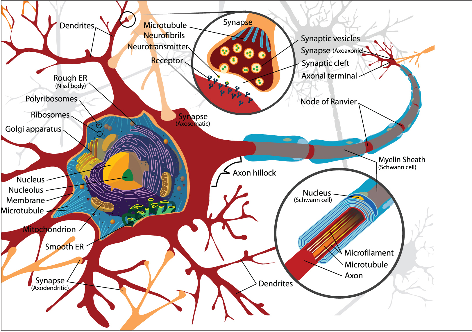
This is the neuron, the building block of the nervous system. Neurons come in many shapes and sizes, but most have some basic features. The cell body contains structures such as the nucleus. Dendrites, the arms extending from the cell body, receive signals from other neurons at junctions called synapses. The neuron sends signals via the axon, a long cable that ends with the axon terminals. The axon terminals release chemical messengers called neurotransmitters.
NEURONS AND GLIA
 The functional unit of neural circuits and networks is the neuron, a specialized cell that can transmit electrical signals to other nerve cells, muscles, or glands. Neurons come in a broad range of shapes and sizes, but all of them have a cell body, dendrites, and an axon. The cell body, also called the soma, contains the neuron’s nucleus and most of its cytoplasm, along with molecular machinery for building and transporting proteins critical to the cell’s function. Dendrites are branched projections that extend from the cell body and collect incoming signals from other neurons. The neuron’s electrical signals travel down its axon — another extension from the cell body that may branch before ending in axon terminals, where the signal is passed across a synapse to other cells. In some neurons, axons are only a fraction of a centimeter long; in others, they may extend more than a meter.
Neurons are associated with support cells called glia. Neuroscientists have long believed that glia outnumber neurons by 10:1 (or more). However, recent investigations suggest that in some regions of the brains of humans and other primates, that ratio is closer to 1:1. However, the ratio of glia to neuron from region to region varies considerably. The central nervous system contains four main types of glial cells: astrocytes, microglia, ependymal cells, and oligodendrocytes. Astrocytes form a network inside the brain that regulates ion concentrations around neurons, provides them with nutrients, and helps regulate the formation of new connections between neurons. Microglia are the main “immune cells” of the brain. They function mainly as phagocytes — helping protect the brain from infections and cellular damage — but can also regulate the formation of new neuronal connections. Ependymal cells make the cerebrospinal fluid that cushions the brain inside the skull, and oligodendrocytes improve neuron function by wrapping axons in a fatty sheath called myelin.
Ion Channels and Action Potentials
Ions are electrically charged atoms that can only cross a neuron’s cell membrane through tunnel-like proteins called ion channels. These tunnel-like proteins act like gates, allowing some ions to enter or leave the cell, but keeping others out. Ions that enter or leave the cell change the voltage difference across the membrane. This change in voltage influences the neuron’s likelihood of generating an electrical signal.
In mammals, the voltage difference across the membrane of a resting neuron is around -70 millivolts (mV), more negative inside the cell than on its outer surface. That membrane potential is affected by signals arriving from other neurons in its circuit, which can make the membrane potential less negative (depolarized) or more negative (hyperpolarized) by opening ion channels in the dendrites. If the sum of all the signals at the dendrites rises to match the membrane’s threshold voltage, a series of voltage-sensitive ion channels opens automatically, triggering an electrical impulse called an action potential, which moves down the axon towards the next neuron in the circuit.
SYNAPSES AND NEUROTRANSMISSION
Signals are passed from one neuron to the next at junctions called synapses. In most circuits, a synapse includes the end of an axon, the dendrite of an adjacent neuron, and a space between the two called the synaptic cleft. Amazingly, this separation between neurons was only verified (by electron microscopy) in the 1950s. The cleft is wide enough that electrical signals can’t directly impact the next neuron; rather, chemical signals called neurotransmitters cross the synapse. This process is called neurotransmission.
Ferreira, et al. The Journal of Neuroscience, 2015.
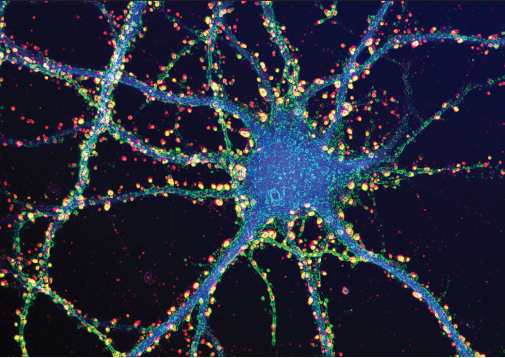
Dendrites — the arms extending from a neuron’s cell body — receive information from other neurons at sites called synapses. Each dendrite can have thousands of synapses, which together form complex circuits that govern brain function. The synapses on this mouse neuron are labeled in yellow and red.
When an action potential arrives at the axon terminal, the voltage change triggers ion channels in the membrane to open, which lets calcium ions flow into the cell. When the calcium ions bind to packages of neurotransmitter molecules called synaptic vesicles, the vesicles fuse with the cell membrane at the axon terminal and empty their contents into the synaptic cleft. Afterwards, pieces of axon terminal membrane cycle back into the soma as new vesicles, which are refilled with neurotransmitter molecules.
Many substances act as nerotransmitters, including amino acids, gases, small organic chemicals, and short peptides. Neurons can synthesize small non-peptides like dopamine or acetylcholine inside the axon terminal. But an axon terminal doesn’t contain the molecular machinery for building proteins, so peptide-based neurotransmitters are built in the ribosome-rich space of the cell body. Vesicles containing neurotransmitter “cargos” bud off from the wall of the Golgi apparatus — the cell’s protein-packaging organelle — then bind to proteins called kinesins that work their way down the axon along microtubules, filamentous parts of the cellular skeleton.
Many different molecules act as neurotransmitters, and each one fits into specific receptors like a key fits a lock.
After neurotransmitters are released from an axon terminal, they drift across the synaptic cleft until they reach the outer surface of the dendrite, a region that looks thick or dense in highly magnified images. This region, the postsynaptic density, has a high concentration of neurotransmitter receptors. Many different molecules act as neurotransmitters, and each one fits into specific receptors like a key fits a lock. Receptors are linked to ion channels in such a way that, when neurotransmitter molecules dock on their receptors, they open those channels, altering the voltage across the postsynaptic membrane. Local glial cells (astrocytes) mop up any excess neurotransmitters at the synapse. This process prevents them from continuously activating receptors.
There are two broad types of receptors on the postsynaptic membrane. In an ionotropic receptor, a neurotransmitter binds directly to part of an ion channel. The channel is normally closed; the receptor protein changes its shape when the neurotransmitter attaches, widening the tunnel in the center of the ion channel so that ions can move through. Metabotropic receptors are more complex. The receptor and the ion channel are different proteins located at a distance from one another, but they are linked by a cascade of biochemical steps that are triggered when a neurotransmitter binds to the receptor. This response is less rapid and activates a series of events inside the postsynaptic cell. The result may be opening an ion channel some distance away or activating other intracellular molecules.
Neurotransmitter molecules only bind to their receptors for a short time. Once they detach, the ion channels return to their resting state and stop altering the charge across their membrane. The neurotransmitters are either broken down or reabsorbed by the axon terminal in a process called reuptake.
The excitatory and inhibitory neurons described above can be identified by the specific neurotransmitters that they make. Excitatory neurons make neurotransmitters that open ion channels that depolarize the dendrite’s membrane; inhibitory neurons make neurotransmitters that hyperpolarize it. The brain’s most common excitatory neurotransmitter is glutamate; the brain’s most common inhibitory neurotransmitter is gamma-aminobutyric acid (GABA).
Glutamate is an amino acid used as a neurotransmitter by approximately half the excitatory synapses in the brain. It can bind to several types of ionotropic receptors; the most important of these are AMPA receptors and NMDA receptors. When activated, the action of AMPA receptors is fast and brief; NMDA receptors activate more slowly, particularly in response to waves of multiple action potentials. Interactions between these receptors appear to be important in learning and memory.
GABA is the brain’s most important inhibitory neurotransmitter. It binds to two groups of receptors; one group is ionotropic, the other metabotropic. Ionotropic GABA receptors have ion channels that let negatively charged chloride ions enter the cell. Metabotropic GABA receptors open ion channels that release potassium ions. In both instances, ion movement pushes membrane potential downward and inhibits a neuron from firing.
RECEPTORS AND MOLECULAR SIGNALING
Neurons have receptors for many molecules that can change the way they function. These molecules include hormones, which send the brain specific cues about the condition and activity of distant tissues in the body; neuromodulators such as the endocannabinoids, cannabis-like chemicals that seem to suppress neurotransmitter release; and prostaglandins, small lipids that change the brain’s response (increasing pain sensitivity) to pain and inflammation.
Individual neurons have receptors for different subsets of hormones and neuromodulators. In each case, these molecules are signals that trigger a series of chemical reactions inside the cell. The process starts when one of these molecules binds to its specific receptor. If the receptor is on the surface of the cell, the bound molecule changes the receptor’s shape across the cell membrane and starts a chain of intracellular reactions. This signal transduction pathway ultimately modifies neuronal function, either by shifting the cell’s ion balance or by changing the activity of specific enzymes.
If a molecule can diffuse through the cell membrane — as occurs with steroid hormones like estradiol or cortisol — its receptor might be a protein inside the neuron’s soma. When the hormone binds to its receptor, the complex can transform into a transcription factor that is capable of entering the cell nucleus, binding to specific genes and changing their activity.
NEURONS, GENES, AND GENE EXPRESSION
By this point, it should be clear that neurons inside the brain can differ in appearance and function. They can produce different types of neurotransmitters, determining whether their signals have excitatory or inhibitory effects in their circuits. They can have different assortments of neurotransmitter receptors, determining the cells’ sensitivity to the effects of specific neurotransmitters. And, in their cell membranes, neurons possess different combinations of receptors capable of detecting neuromodulators that influence neuronal behavior — for example, hormones such as vasopressin, estradiol, or cortisol.
All cells in your body, including neurons, contain the same DNA housing the same genes. Differences among your neurons result from differences in which genes direct cellular activities, a process called gene expression. Each cell (or cell type) builds proteins from a slightly different subset of genes in its genetic code, the same way different children will build different structures from the same starting set of Lego blocks.
The mechanisms causing neurons to express some genes and not others are currently an area of intense research. Many of these mechanisms depend on chemical changes to chromatin, the complex of protein and DNA that compactly packages the long DNA molecule inside the nucleus. Genes that a cell is using to build proteins need to be accessible and are associated with open, unfolded chromatin, while unexpressed genes are typically in tightly packed regions. Chemical changes that tighten or spread out chromatin complexes can, respectively, shut down or activate the genes on that segment of DNA. These changes are reversible, giving neurons flexibility to alter the genes they express in response to hormonal cues and environmental changes.
The genes that affect neuron structure and function can also differ between individuals. Gene variants or alleles reflect differences in the nucleotide sequences that make up a gene. While different alleles code for forms of the same protein, the variants can produce structural differences that affect their function. An allele might code for a version of an enzyme that is less effective than the usual version, and specific alleles of some genes can even cause neurological diseases. For example, Tay-Sachs disease, a fatal degenerative neurological condition, is caused by mutations in a gene that codes for part of a fat-metabolizing enzyme called beta-hexosaminidase A. Because the variant enzyme is poor at breaking down specific fats, these build up in neurons and become toxic. There are many cases where small changes in genetic sequence affect how our brain can function, and in the next 10 years — with our capacity to sequence a person’s entire genome now possible — we will be able to move much closer to understanding the genetic basis of brain disorders. 